During mitosis DNA is duplicated, resulting in two identical chromosomes. These are held together at a special part of the chromosome, called the centromere.
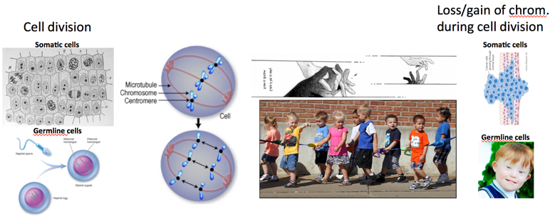
On the top of the centromere, a protein megacomplex – the kinetochore – forms to connect duplicated chromosomes to microtubules that emanate from opposite poles of the dividing cell. Only when all the chromosomes attach correctly to microtubules, are the sister chromatids separated. These then travel to the poles of the dividing cell.
Making sure that each chromosome is attached correctly is very challenging – it can be likened to making sure that 23 pairs of kindergarteners are all holding hands whilst being dragged in opposite directions and, at the same time, managing to form a straight line. The accuracy with which cells control this complex process, which happens at least a billion times a day in our organism, is intriguing.
Mistakes resulting in inaccurate chromosome segregation are either detrimental, or typical of cancer cells. Furthermore, in cases of unequal division in germ cells, the results are congenital genomic disorders, such as Down syndrome.
Thus, a better understanding of the molecular mechanisms that guard the genome during these numerous cell divisions also means a better understanding of the basics of genetics and heredity itself. However, further research in this field also holds promise to initiate new therapies for health conditions.
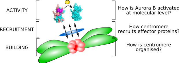
Our research focuses on centromeres. We want to understand:
1) What are the structural determinants of centromere formation and maintenance?
2) How does the centromere recruit major effector proteins in mitosis?
3) What is the molecular basis for the activation of central mitotic kinase, Aurora B?
Our Lab is equipped with state-of-the-art instrumentation for structural biology and mass spectroscopy. We also collaborate closely with experts in yeast genetics and cell cycle regulation.