I min forskning er jeg interessert i å identifisere mekanismene som ligger til grunn for hjernens fantastiske evne til å prosessere sanseinformasjon og lagre erfaringer i nevrale nettverk. Vår forskningsgruppe jobber in vivo med elektrofysiologiske registreringer av store populasjoner av nerveceller. For å se sammenhengen mellom nervecellenes struktur og funksjon benytter vi genteknologi og to-foton laser-scanning mikroskopi.
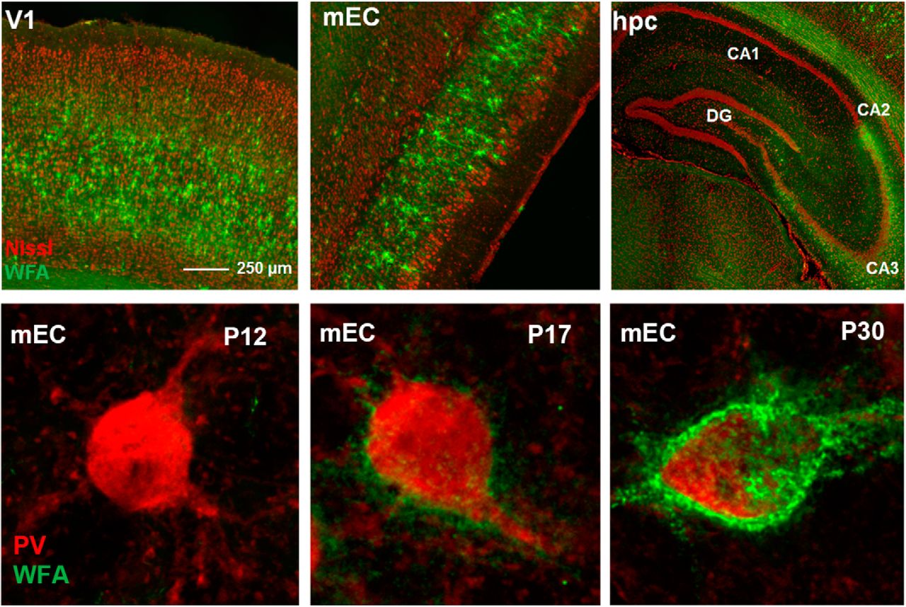
Inhibitoriske nevroner (PV+) i rødt, perinevrale nett (grønt) i visuell cortex, medial entorhinal cortex (MEC) og hippocampus. Nederste rad viser dannelsen av nettene rundt synapsene i løpet av kritisk periode.
Undervisning
Mesteparten av min undervisning er i nevrofysiologi på medisinstudiet, Modul 2.
The medial entorhinal cortex (MEC) is pivotal in spatial computations and episodic memory. In particular, an animal’s position can be decoded from the activity of entorhinal grid cells. However, it remains elusive whether MEC could play a more general role in different types of associative learning and how the representations develop during the learning process. It has been shown that the postrhinal cortex (POR), which is directly connected to MEC, integrates visual stimuli with salient outcomes. Here, we use a non-spatial visual association task to investigate whether MEC neurons represent low-level visual cues during learning. Using a Go/NoGo visual association task, we recorded neural activity in MEC and POR throughout the learning phase as mice associated drifting gratings with rewarded, aversive, or neutral outcomes. Our findings reveal that the neural tuning curves in both the POR and MEC change with the learning of the task. From the start of training, the POR neurons exhibited response tuning to the visual cues, and the tuning was stable to cue orientations during learning. In contrast, MEC neurons did not initially respond very strongly to visual cues but developed a robust tuning toward the rewarded trials. While the MEC’s representation of visual information was limited, it encoded other task elements. A large fraction of the neurons formed distinct functional clusters that were either activated or suppressed by reward-related behavior. Remarkably, these clusters segregated anatomically in MEC and maintained strong within-cluster correlations before and after training. Notably, although the same functional clusters were apparent in the POR, they did not show any anatomical structure as in the MEC. Task reversal induced significant changes in network responses across both regions, with a decrease in overall task-responsive neurons but a slight increase in stimulus representation. Strikingly, information about the choice to lick emerged with learning in both brain areas, and most significantly within the functional cell clusters representing reward consumption and plus-cue stimulus. Our results demonstrate that although neurons in MEC and POR develop behavior-modulated tuning during learning of a non-spatial visual association task, the MEC exhibits stronger within-cluster correlations and anatomical organization. Conversely, the POR population exhibits less structural organization and more specific stimulus-tuning, which is reflective of being a higher visual association area. Our findings reveal that the MEC can encode task– and behavior-related variables beyond spatial information.
Understanding the neural mechanisms of sleep is critical for advancing treatments for neurodegenerative diseases. To investigate these mechanisms, long-term electrophysiological studies are conducted in animal models, where brain activity can be monitored continuously under controlled conditions. However, electrophysiological recordings from freely moving animals are typically performed using a tethered setup, which imposes constraints on natural behaviour. As a first step towards a miniaturised, wireless neural implant, we present a low-power system that enables direct control of the Intan RHD2132 electrophysiology chip using a custom FPGA-based stack. A light-weight yet robust implementation ensures efficient operation while maintaining the necessary flexibility for future integration into a compact, wireless system. The FPGA handles real-time signal processing and data transmission, with support for closed-loop stimulation and onboard data compression. Our system achieves continuous multi-channel recording at 20 kHz, with real-time wireless streaming and long-term data storage. The hardware design prioritises low power consumption, with the aim of enabling extended operation for uninterrupted sleep monitoring over 24 hoursClinical Relevance— This technology enables long-term, high-resolution sleep monitoring, which is essential for understanding the role of sleep in neurodegenerative disorders such as Alzheimer’s and Parkinson’s. By providing continuous, naturalistic brain recordings, this system may help identify early electrophysiological biomarkers of cognitive decline, with potential translational and therapeutics applications.
Perineuronal nets (PNNs) are a condensed form of extracellular matrix primarily found around parvalbumin-expressing (PV+) interneurons. The postnatal maturation of PV+ neurons is accompanied with the formation of PNNs and reduced plasticity. Alterations in PNN and PV+ neuron function have been described for mental disorders such as schizophrenia and autism. The formation of PNNs is highly dependent on aggrecan, a proteoglycan encoded by the ACAN gene, but it remains unknown if it is produced by the PV+ neurons themselves. Thus, we established a knockout (KO) mouse model (ACANflx/PVcre) and an adeno-associated virus to specifically eliminate aggrecan production from PV+ neurons, in the germline or adult animals, respectively. The germline KO (ACANflx/PVcre) eliminated the expression of PNNs labeled by Wisteria floribunda agglutinin (WFA), the most commonly used PNN marker. Surprisingly, electrophysiological properties of PV+ interneurons and ocular dominance plasticity of adult ACANflx/PVcre mice were similar to controls. In contrast, AAV-mediated ACAN knockout in adult mice increased ocular dominance plasticity. Moreover, in vivo Chondroitinase ABC treatment of KO mice resulted in reduced firing rate of PV+ cells and increased frequency of spontaneous excitatory postsynaptic currents (sEPSC), a phenotype associated with chABC treatment of WT animals. These findings suggest that compensatory mechanisms may be activated during development in response to the germline loss of aggrecan. Indeed, qPCR of bulk tissue indicates that other PNN components, including neurocan and tenascin-R, are expressed at higher levels in the KO animals. Finally, behavioral testing revealed that ACANflx/PVcre mice had similar long-term memory as controls in the Morris water maze. However, they employed bolder search strategies during spatial learning and showed lower level of anxiety-related behavior in an open field and zero maze.
Highly salient events activate neurons across various brain regions. During subsequent rest or sleep, the activity patterns of these neurons often correlate with those observed during the preceding experience. Growing evidence suggests that these reactivations play a crucial role in memory consolidation, the process by which experiences are solidified in cortical networks for long-term storage. Here, we use longitudinal two-photon Ca2+ imaging alongside paired LFP recordings in the hippocampus and cortex, to show that targeted manipulation of PV+ inhibitory neurons in the lateral visual cortex after daily training selectively attenuates cue-specific reactivations and learning, with only minute effects on spontaneous activity and no apparent effect on normal function such as visual cue–elicited responses during training. In control mice, reactivations were biased toward salient cues, persisted for hours after training had ended, and the prevalence of reactivations was aligned with the learning process. Overall, our results underscore a crucial role for cortical reactivations in memory consolidation.
Beshkov, Kosio; Fyhn, Marianne; Hafting, Torkel & Einevoll, Gaute
(2024).
Topological structure of population activity in mouse visual cortex encodes densely sampled stimulus rotations.
iScience.
27(4).
doi: 10.1016/j.isci.2024.109370.
Fulltekst i vitenarkivVis sammendrag
The primary visual cortex is one of the most well understood regions supporting the processing involved in sensory computation. Following the popularization of high-density neural recordings, it has been observed that the activity of large neural populations is often constrained to low dimensional manifolds. In this work, we quantify the structure of such neural manifolds in the visual cortex. We do this by analyzing publicly available two-photon optical recordings of mouse primary visual cortex in response to visual stimuli with a densely sampled rotation angle. Using a geodesic metric along with persistent homology, we discover that population activity in response to such stimuli generates a circular manifold, encoding the angle of rotation. Furthermore, we observe that this circular manifold is expressed differently in subpopulations of neurons with differing orientation and direction selectivity. Finally, we discuss some of the obstacles to reliably retrieving the truthful topology generated by a neural population.
Olsen, Elise Reppe; Fjeldstad, Maria Perona; Thon, Sarah Kriebel; Schøyen, Vemund Sigmundson; Lepperød, Mikkel Elle & Sugar, Jørgen Afseth
[Vis alle 8 forfattere av denne artikkelen](2024).
Exploring social and spatial coding in the lateral and medial entorhinal cortex.
Fulltekst i vitenarkiv
Fjeldstad, Maria Perona; Lepperød, Mikkel Elle; Schøyen, Vemund Sigmundson; Christensen, Ane Charlotte; Grødem, Sverre & Fyhn, Marianne
[Vis alle 7 forfattere av denne artikkelen](2024).
Theta modulation of grid cells in the medial entorhinal cortex in a novel environment task.
Fulltekst i vitenarkiv